WHAT IS ANGIOGRAPHY ?
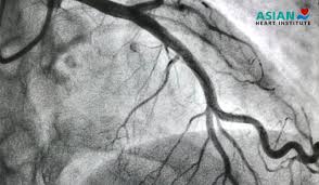
Angiography is a medical imaging technique used to visualize the inside of blood vessels and organs of the body, particularly the arteries and veins, using X-rays and a contrast dye. It helps doctors diagnose and treat various conditions, including narrowed or blocked blood vessels, aneurysms, and structural heart or valve diseases.
Here’s a more detailed explanation:
How it works:
- A thin, flexible tube called a catheter is inserted into a blood vessel, usually in the groin or arm.
- A special dye (contrast agent) is injected through the catheter, making the blood vessels visible on X-ray images.
- X-ray images are taken as the dye flows through the vessels, revealing their structure and any abnormalities.
Types of Angiography:
- Coronary Angiography: Focuses on the blood vessels of the heart (coronary arteries).
- CT Angiography (CTA): Uses computed tomography (CT) and contrast dye to visualize blood vessels.
- Magnetic Resonance Angiography (MRA): Uses magnetic resonance imaging (MRI) and contrast dye to visualize blood vessels.
Why it’s used:
- Diagnosis: To identify blockages, aneurysms, or other abnormalities in blood vessels.
- Treatment: As a first step in procedures to open blocked arteries (like angioplasty).
- Monitoring: To assess the effectiveness of treatments for vascular conditions.
Potential risks:
Bleeding or bruising at the catheter insertion site, Allergic reaction to the contrast dye, and Damage to blood vessels.
In summary, angiography is a valuable tool for visualizing blood vessels, aiding in the diagnosis and treatment of various conditions affecting the circulatory system.


Post Comment